Gene Therapy Helped Adult Dogs’ Retinas Rebuild
Researchers expected better vision. They did not expect mature retinas to rebuild some of their damaged wiring.
Key takeaways
- The dogs carried a faulty CaBP4 gene that disrupted communication between retinal cells.
- Treated eyes regained electrical responses and performed far better in dim-light navigation tests.
- The mature retinas physically rebuilt some damaged wiring instead of merely slowing further decline.
- The treatment is experimental, limited to one rare inherited disorder and unavailable through routine veterinary care.
For decades, a common assumption in neuroscience has been that once nerve connections in an adult mammal are damaged, they mostly stay that way. A new study in dogs adds a notable piece of evidence to the other side of that argument.
Researchers at Michigan State University’s College of Veterinary Medicine reported that a single-dose gene therapy did more than slow an inherited form of blindness in dogs. In the treated part of the eye, the retina reorganized and rebuilt connections between light-sensing cells and the nerve cells that pass their signals on, and the dogs’ vision in dim light improved.
The paper, led by Billie Beckwith-Cohen and Simon Petersen-Jones, appears in Molecular Therapy Advances. It was published online in June 2026 and appears in the journal’s September 10 issue; the university highlighted it again in late September.
What condition did the dogs have?
The dogs came from a Michigan State research colony bred from a whippet sire and dam that originated in Brazil. They carry a naturally occurring mutation in a gene called CaBP4, which makes a calcium-binding protein used at the retina’s first synapse—the junction where photoreceptors, rods and cones, hand signals to the next layer of cells.
Without working CaBP4, that handoff fails. The researchers found that the dogs’ synaptic “ribbons,” tiny structures that help photoreceptors release their signals, stayed short and immature. The outer plexiform layer, where these connections live, was thin and disorganized. Standard eye-function tests showed a characteristic abnormal pattern, and the dogs struggled to see in dim light. Over time, the light-sensing cells also began to die.
In people, CaBP4 mutations cause visual problems from childhood. The authors say the most fitting name for the condition is “congenital cone-rod synaptic disorder.” MSU describes it as a rare form of inherited blindness.
What did the researchers do?
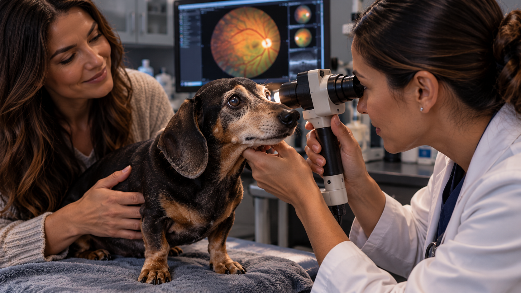
The team used a modified virus widely used in gene therapy—an adeno-associated virus, or AAV—to deliver a working copy of the CaBP4 gene. Surgeons injected it under the retina, creating a small treated “bleb” rather than covering the entire retina. In most dogs, one eye was treated and the other served as an untreated comparison.
The main treatment group averaged about a year old at the time of treatment. Two dogs were treated at 28 months, when considerable photoreceptor loss had already occurred. Some dogs were followed for more than two years, and the authors report that structural recovery was maintained for up to three years after treatment.
What happened to vision?
Electroretinography, which records the retina’s electrical response to flashes of light, showed that signals missing before treatment returned in treated eyes, including in the two dogs treated later in the disease.
The team also tested practical vision. In a four-choice exit device, dogs had to find the one open tunnel at different light levels. Treated eyes chose correctly more often and exited faster than untreated eyes, and performance was comparable with unaffected dogs. On an obstacle course in dim light, treated eyes finished faster with fewer collisions.
Under bright light, the improvement was not statistically significant. The authors attribute that result to the dogs still having some residual daytime vision at the ages tested, leaving less room to show a difference.
Why the structural changes surprised researchers
Functional improvement was the expected goal. What stood out was physical repair in retinas that were already mature.
- Ribbons grew up. In treated regions, synaptic ribbons lengthened to roughly normal size—about 604 nanometers, compared with about 642 in unaffected dogs and about 227 in young untreated affected dogs. Untreated regions stayed short.
- The connection layer thickened. The outer plexiform layer expanded to about 6.8 micrometers in treated regions, versus about 1.7 in untreated regions and 8.2 in normal dogs. It grew beyond its thickness before treatment, meaning cell processes extended to rebuild the layer.
- Partner proteins returned. GPR179, a key protein on the receiving side of the synapse, recovered to levels comparable with normal dogs in treated areas. Imaging also showed a restored network of connections between cones and the cells they signal.
“Not only were new components added, but pre-existing abnormalities were repaired,” Beckwith-Cohen said in the university’s summary.
She described the mutation as “a typo in a blueprint” and the therapy as supplying corrected instructions.
Why dogs matter to this research
Much eye research uses rodents, but dogs have larger eyes, well-developed vision and a retinal region dense in cones, which the MSU team says makes them closer to people in important ways. Because these dogs developed the condition naturally rather than through engineered mutations, the researchers say the model better represents how the disease behaves.
MSU notes that canine gene-therapy research at the college has a long track record of informing human clinical work. Still, a successful result in one naturally occurring dog model is a research milestone—not proof that the same approach will work, or be safe, in people or in other diseases.
What this does not mean
- It is not a cure for common vision loss. The study covered one rare genetic disorder. It says nothing about reversing cataracts, glaucoma, eye infections, injuries or age-related vision changes.
- It is not a routine veterinary treatment. This was experimental surgery using a research gene-therapy product.
- Recovery was partial and local. Only the injected area of the retina was treated, and the numbers of dogs were small, all from a single colony.
- Human benefit is not established. The authors say the work “lays the foundation” for clinical studies in the analogous human condition. That is a next step, not a result.
The broader takeaway is the one that has researchers talking: when the missing piece was restored, adult retinal wiring showed more capacity to reorganize than many expected. The researchers suggest that understanding which kinds of damage can be reversed may matter for other inherited retinal diseases and possibly beyond the eye.
If your dog seems hesitant in dim light, bumps into objects or shows any change in the eyes, the practical step is unchanged: see your veterinarian, who can refer you to a veterinary ophthalmologist if needed.
Sources and further reading
- Beckwith-Cohen B, Sun K, Occelli LM, et al. Gene therapy induces synaptic ribbon maturation, synaptogenesis and vision recovery in an adult dog model of retinal degeneration. Molecular Therapy Advances, 2026.
- Michigan State University College of Veterinary Medicine — MSU Researchers Show Adult Retinas Can Repair Themselves
- MSUToday — Researchers Show Adult Retinas Can Repair Themselves
This article provides general educational information and is not a substitute for veterinary advice, diagnosis or treatment.
About the author
Stephen Gill
Stephen Gill is the founder of Inspired by Tyson and editor of Daily Doggy News. His work covers dog health, behavior, safety, research and everyday life with dogs.
Read Stephen Gill’s full author profile